Cartilage- Functions, Structure, and Characteristics
Introduction
Cartilage is a type of connective tissue that is found throughout the body, from the nose to the ends of our fingers. Cartilage can be rigid and stiff or soft and flexible depending on its location in the body.
This article will examine cartilage definitions, structure, functions, and characteristics to help you better understand how it works! However, should you chose to skip this guide due to reasons such as a busy schedule, our professional writers for hire are ready to cover you by doing that assignment for you. Just place an order and leave it to us.

Cartilage Structure and the Main Components?
The cartilage is a dense, sponge-like matrix of collagen fibers located throughout the body. Cartilage is formed of tiny cells called chondrocytes and is surrounded by a thin, transparent membrane. It has an appearance similar to that of the brain and the spinal cord.
Cartilage is a type of connective tissue that gives strength and elasticity to joint bones and protects the bones. It is found everywhere in the body, and its characteristics depend on the specific location.
Chondroitin sulfate is a major component of cartilage which is a glycosaminoglycan. It’s an essential part that composes the matrix of collagen fibers and provides the cartilage with its elasticity and protects the bone.
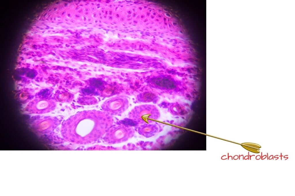
An extracellular matrix binds chondrocytes together and allows the cartilage to be flexible and elastic. Chondroblasts are the other specialized cells of the cartilage tissue found in the cartilage matrix. They are responsible for producing new cells and for rebuilding damaged tissue.
You may be interested in Chromatins
The Main Components of Cartilage
- Fibroblasts
- Chondroblasts
- Chondrocytes
- The Extracellular matrix
Fibroblasts are part of all connective tissues, and they produce the collagen fibers that make up most of the cartilage. They are responsible for producing collagen protein and giving cartilage its elasticity.
Chondroblasts are the cells responsible for replenishing and maintaining cartilage. Chondrocytes are specialized types of fibroblasts that produce chondroitin sulfate.
The extracellular matrix, which makes up the majority of the cartilage, is a fluid-like, healthy environment. It provides the joint with its elasticity and strength while also creating a protective cushion for the bone.
The matrix comprises collagen, fibronectin, lipids, proteoglycans, and the cells; these make up bone cartilage. It supports the cells that produce it while providing a medium for the exchange of nutrients and waste products between the cells and their surrounding environment.
The caoutchouc is a type of cartilage that has the appearance and elasticity of rubber. It’s found in areas like the ear, nose, and larynx.
The cartilage that is located in the eardrum, called the tympanic membrane, is an example of elastic cartilage. It is sensitive to heat and cold because the extracellular matrix does not have a high tolerance for temperature changes. This explains why the cartilage is damaged when exposed to extreme heat or cold.
The Types of Cartilage
There are three different types of cartilage:
- Elastic Cartilage
- Fibrocartilage
- Hyaline Cartilage
A) Elastic Cartilage
Elastic cartilage is found in the larynx, trachea, and bronchial tubes, where it provides elasticity. It is a connective tissue that consists of collagen and elastin fibers that allow for flexibility.
Elastic cartilage is the type of cartilage most often found in the body and provides a free-flowing, flexible environment for bones. This connective tissue is highly flexible hence withstands bending pressure.
This type of cartilage provides elasticity to the bones and protects them from being crushed. It also helps to maintain the shape of the external ear. In the human body, elastic cartilage is also found in the epiglottis, providing elasticity in the trachea.
B) Fibrocartilage
Fibrocartilage is a dense connective tissue with a higher concentration of collagen fibers than hyaline cartilage and is found in the ear, nose, and ribs.
The fibrocartilaginous type of cartilage is the most resistant to pressure, and it has a heavy collagen fiber matrix. It also protects from injury, shock absorption, and stability for the joint.
Of the three types of cartilage, fibrocartilage is the only one that contains both types I and Type II cartilage. It is found in the menisci, the discs between the joints, and ligaments.
C) Hyaline Cartilage
Hyaline cartilage is located in places that are exposed to a lot of pressure and stress, such as the joints, rib cage, and larynx.
The hyaline cartilage is also found in the nose and ears; it is soft, spongy, and flexible. It is also found in the pubic symphysis, where it helps to provide elasticity.
This type of cartilage contains a high concentration of collagen fibers which provide flexibility. It is also very light because of the high water content, making it more elastic and less dense than fibrocartilage.
The elastic cartilage hyaline also contains a type of proteoglycan, chondroitin sulfate. This is the most common type of cartilage in humans. It provides flexibility and elasticity while also being incredibly lightweight.
The bones of an embryo form as hyaline cartilage then later develop into bones. This fact makes hyaline very crucial in fetal development.
Characteristics of The Three Types of Cartilage
The different types of cartilage have both similarities and differences in structure, functions, and locations:

Similarities between the Hyaline Cartilage, Fibrocartilage, and Elastic Cartilage
- Both the hyaline cartilage and elastic cartilage are made of type I collagen fibers. They also have a thicker extracellular matrix than fibrocartilage, and they have high water content.
- The three types of cartilage have a similar function: they provide structural support and protection for the body. Also, they have the same kind of cells called chondrocytes.
The Differences
- The elastic cartilage has more elastin fibers than the hyaline cartilage, which makes it more flexible.
- The elastic cartilage is also connected to muscles and bones by tendons, ligaments, and joint capsules.
- Fibrocartilage is the stiffest among all three types of cartilage because it has a higher concentration of collagen fibers.
- Fibrocartilage is the least elastic among all three types, and it also has less type I collagen than hyaline cartilage.
- The function of the fibrocartilaginous type of cartilage is to provide protection and stability for joints while also protecting them from injury, shock absorption, and providing stiffness.
- The hyaline cartilage is mainly in the nose, ear, pubic symphysis (joint), and larynx. It is also found in the rib cage, joints, nose, ear, and the pubic symphysis (joint). Here, it provides elasticity, protection, and stability.
- The fibrocartilage is found in the meniscus (cartilaginous discs) between the joints and in ligaments. It provides protection, stability, and stiffness to the joint while also providing shock absorption.
- The elastic cartilage is located in the pubic symphysis (joint). It provides stability, protection, and flexibility.
Describe the Process of Cartilage Formation
The process of cartilage formation is called chondrification. The two significant factors that affect cartilage formation are:
- Type I collagen fibers provide elasticity and protection to the body.
- Type II collagen fibers provide stability to the joint.
The cartilage is made up of cells called chondrocytes which produce the cartilage’s extracellular matrix.
The matrix contains proteoglycans, glycoprotein, and water which protect the body. The cells also produce more type II collagen fibers that provide stability to the joint.
Another factor is mechanical stress, which causes chondrocytes to produce more proteoglycans and water, making the cartilage stronger, thicker, and stiffer.
During formation, the cartilage cells are in a resting phase before going back into a proliferative stage. The chondrocytes produce type I collagen fibers that provide elasticity and protection to the body.
How Does Cartilage Grow?
Cartilage tissue growth depends on the increased size or mass of the cartilage. It does not grow in length or width as it is a connective tissue and lacks joint structures.
The growth of cartilage is caused by the increase of water and proteoglycans in the extracellular matrix. It also involves the addition of hyaluronic acid that provides lubricant for joints.
The cell size also determines how fast cartilage grows. Cells in the growth phase will produce more proteoglycans and water that increase the size of cartilage.
You may be interested in Parts of a Small Intestine
Cartilage Repair
There are many different ways that cartilage can be repaired. Some of the methods include:
Rheumatoid Arthritis Treatment
RAT is a type of treatment that helps to reduce inflammation and slow the progression of joint damage caused by rheumatoid arthritis.
Cartilage Transplant
Cartilage transplant is a treatment that involves replacing the cartilaginous tissue with healthy tissues from other parts of the body.
Cartilage Augmentation
It is a treatment done before the cartilage has degenerated. It can be either a temporary or permanent method.
Cartilage Restoration
This treatment is done after the cartilage has degenerated. It can be either a temporary or permanent method.
Cartilage Stripping
It refers to the treatment done if the cartilage has degenerated and cannot restore cartilage.
Cartilage Induced Osteogenesis
Induced Osteogenesis is a treatment done if the cartilage has degenerated and it’s not possible to restore the cartilage. It’s done by transplanting bone marrow cells or stem cells and providing them with a milieu that will induce the growth of new cartilage.
You may be interested in the Endomembrane System
Summary
Cartilage is a type of connective tissue which provides support to joints. It is mainly of water, collagen fibers, and proteoglycans (proteins containing sugar molecules). In general, cartilage has the following functions and characteristics: it’s rigid but flexible; it cushions surfaces so they don’t touch directly; its cells regenerate faster when damaged or worn away.
There are three main types of cartilage in your body – articular cartilage, intervertebral discs, and costal cartilages—research more about the structure of different types of cartilage to understand how the human body works.